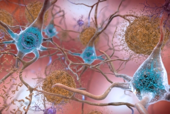
Scientists at UC Santa Barbara and Northwestern University have created the first synthetic fragment of tau protein that acts like a prion. The “mini prion” folds and stacks into strands, or fibrils, of misfolded tau proteins, which then transmit their abnormally folded shape to other normal tau proteins. Misfolded, prion-like proteins drive the progression of tauopathies, a group of neurodegenerative diseases — including Alzheimer’s disease — characterized by the abnormal accumulation of misfolded tau protein in the brain. By studying a minimal synthetic version of the full-length human tau, scientists can better recreate the fibril structure containing misfolded tau proteins. This potentially could lead to targeted tools for diagnosis and therapy that are much needed for neurodegenerative diseases.
While developing the synthetic protein, the scientists also uncovered new insights into the role of water around the protein surface that guides the misfolding process. A mutation commonly used to model tau-related diseases subtly changes the dynamic structure of water in the environment immediately surrounding the tau protein, the researchers found. This altered water structure influences the protein’s ability to adopt its abnormal shape. The study was published on April 28 in The Proceedings of that National Academy of Sciences.
“The scope of neurodegenerative diseases involving the protein tau is particularly broad,” said Songi Han, who led the study. “It encompasses chronic traumatic encephalopathy, which is found in football players after head trauma, corticobasal degeneration or progressive supernuclear palsy. Creating self-propagating tau fragments that can recreate the fibril structure and misfolding that is unique to each tauopathy disease is a crucial step forward in our ability to understand and model these complex diseases.”
Han, a professor of chemistry at Northwestern, started the work while a professor of chemical engineering and chemistry at UCSB. Michael Vigers, who received his PhD in chemical engineering at UCSB and now works as an associate researcher at Northwestern, was first author of the paper. UCSB coauthors included chemical engineering professor M. Scott Shell, as well as molecular, cellular, and developmental biology professor Kenneth S. Kosik and chemistry professor Joan-Emma Shea. The work also was made possible by several students and postdoctoral fellows, including UCSB chemical engineering PhD student Samuel Lobo and postdoctoral scholar Saeed Najafi.
A chain-reaction of misfolding
In many neurodegenerative diseases, proteins misfold and clump together into harmful, highly ordered fibrils, which ultimately damage brain health but are difficult to diagnose. When a normal protein encounters the pathological tau fibrils, the normal protein changes shape to match the misfolded form. This process leads to a chain reaction, where more and more proteins transform into the misfolded, aggregation-prone state. Although this behavior is prion-like, it does not involve actual prions, which can spread contagious diseases from person to person.
Using cryogenic electron microscopy (cryo-EM), researchers solved the structure of the fibrils from samples of brain tissue. Although pinpointing the structure was a significant breakthrough, brain samples can only be obtained after a patient dies. Despite dramatic progress and intense interest in this area, final diagnosis of tau-related neurodegenerative diseases is only possible after death.
“When people start to show signs of neurodegenerative disease, it is not diagnosed today with a biomarker,” Han said. “Physicians determine the diagnosis by administering a patient survey and by examining a collection of symptoms, like sleep patterns and memory. The bottleneck is the reliable generation of tau fibrils that recreate the critical and unique disease hallmarks to serve as targets for developing diagnostic strategies.”
A simplified model
To meet the current challenge, the researchers sought to develop a synthetic, prion-like tau protein. Instead of recreating the entire length of the protein, which is long and unwieldy, Han’s team aimed to pinpoint the shortest piece of tau that could still adopt a misfolded shape and form disease-like fibrils.
Ultimately, they focused on a short segment of tau, dubbed jR2R3, which is just 19 amino acid segments in length. The segment contains a mutation called P301L, commonly found in many diseases. The researchers found this short peptide could form the harmful fibrils, which are the hallmark of these diseases, and act as a “seed” to template the misfolding and aggregation of full-length tau proteins.
“We made a mini version that is easier to control,” Han said. “But it does all the same things that the full-length version does. It does the seeding, causing normal tau protein to misfold and join the fibrils.”
In this work, UCSB professors Shell and Shea, as well as Najafi and Lobo, used atomistically resolved simulations to create a detailed molecular picture of the interactions that underlie the aggregation of the tau fragment. They discovered that the important point-mutant P301L, key to the ability of the jR2R3 construct to aggregate, at the molecular level shifts this protein from compact, aggregation-resistant conformations to more open aggregation-competent shapes. The finding suggests the mutation plays a crucial role in directing the protein to misfold.
After hypothesizing that something must be holding the misfolded proteins together, the simulations identified water surrounding a protein, particularly the water molecules, as playing a crucial role in protein folding and aggregation. The P301L mutation appears to directly change the structure of the tau protein as well as change the behavior of water molecules around it.
“Water is a fluid molecule, but it still has structure,” Han said. “The mutation in the peptide might lead to a more structured arrangement of water molecules around the mutation site. This structured water influences how the peptide interacts with other molecules, pinning them together.”
In other words, organized water pins the proteins together, enabling individual strands to fold together into a neat stack. Then, using their prion-like behavior, the fibrils recruit other proteins to misfold and join the stack.
"This has been a wonderfully integrated team between experiment and theory that has allowed us to translate the capabilities of molecular simulations to create hypotheses for experiments, and then the experiments come back to the simulations with new ideas and directions to explore,” explained Shell, the John Meyers Founders Chair and vice chair of UCSB’s Chemical Engineering Department. “The result has been a new model of tau aggregation with a deep understanding of molecular mechanisms and interactions that will make it a powerful tool for understanding and addressing pathological tau aggregation moving forward."
What’s next
The research team is now focused on further characterizing the properties of the synthetic, prion-like proteins. Eventually, they plan to explore potential applications, including the development of new diagnostic and therapeutic approaches for tau-related diseases.
“Once a tau fibril is formed, it doesn’t go away,” Han said. “It will grab naïve tau and fold it into the same shape. It can keep doing this forever and ever. If we can figure out how to block this activity, then we could uncover new therapeutic agents.”
The study was supported by the National Institutes of Health, Deutsche Forschungsgemeinschaft and the W. M. Keck Foundation. The initial writeup about this study's finding was written by Amanda Morris and published in Northwestern Now.